MRI-Based Brain Tumor Segmentation Using Multi-Scale Graph Learning and CNN Boundary Refinement
DOI:
https://doi.org/10.65521/ijacect.v15i1.2391Keywords:
Abstract
Brain tumor segmentation from Magnetic Resonance Imaging (MRI) is essential for early diagnosis and effective treatment planning. However, accurate segmentation remains challenging due to variations in tumor shape, size, intensity, and location across patients. Traditional Convolutional Neural Network (CNN)-based approaches effectively capture local image features but often fail to model long-range dependencies, while graph-based methods capture global structural relationships but may lack precise boundary delineation.
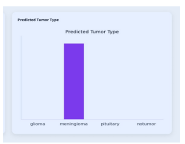
To address these limitations, this work proposes a Multi-Scale Graph-CNN Framework for brain tumor detection, segmentation, and classification. The system supports multiple medical imaging formats, including PNG, NIfTI, and clinical DICOM (.dcm) data. MRI data is preprocessed using normalization and resizing techniques, and multi-scale supervoxels are generated to construct anatomical brain graphs. A Graph Neural Network (GNN) is employed to capture global contextual relationships and produce coarse tumor localization through activation maps. A refinement CNN further enhances segmentation accuracy by generating pixel-level tumor masks. Additionally, a classification CNN predicts tumor types such as glioma, meningioma, pituitary, or no tumor, along with confidence scores.
The proposed system is evaluated using the BraTS dataset and provides comprehensive outputs including tumor probability, activation maps, refined segmentation masks, and classification results. By effectively combining global structural context with fine-grained local details, the framework delivers robust and interpretable results. The integration of DICOM support and an end-to-end interactive interface makes the system a practical and clinically relevant tool to assist radiologists in diagnosis and treatment planning.
Downloads
Published
How to Cite
Issue
Section
License

This work is licensed under a Creative Commons Attribution-NoDerivatives 4.0 International License.